Showing posts with label Tutorials. Show all posts

Saturday, December 18, 2010

What is Multiple Cloning Sites?

A multiple cloning site (MCS) is a section of DNA, usually 20+ restriction sites. These restriction sites are different areas that a gene can be inserted into, and represent complimentary ends of DNA sequences that will adhere to the section of DNA to be inserted.

Restriction sites can have blunt, or 'sticky' ends (like an overhang), and are produced using restriction enzymes that cut the DNA in a specific way. The DNA to be inserted is then cut with the opposing sequence, so that a join can be formed.

MCS are typically found within engineered plasmids (circular double stranded DNA molecules), where they allow sections of inserted DNA be expressed.

An example of this would be the production of Insulin for sufferers of diabetes. The gene for insulin is inserted into a the MCS of a plasmid. The plasmid is then transfected into a bacteria or yeast cell, where the DNA within the plasmid is read, and expressed, leading to the production of insulin.


Few Questions about Multiple Cloning Sites:

In plasmids such as Bluescript with a multiple cloning site in a modified lacZ gene, a cloned insert can be detected by a change of colour from blue to white?

yes, the insert disrupts the lacZ gene and so if you plate the bacteria with x-gal (a substrate to turn blue), the colonies that have the insert will remain white while the colonies that don't will stay blue. lacZ when uninterrupted allows the x-gal to be metabolized and turn blue.

Why is it important that the multiple cloning site of DNA has only one restriction enzyme recognition site? I know that it makes the plasmid linear instead of circular but why does the plasmid need to be linear?

Because if it has two sites you'll end up with two pieces of DNA instead of one piece.

I have cloned my gene of interest in pUC18 at multiple cloning site. can i use this plasmid for over expression?

Yes, you can use it for over expression. Make sure your plasmid has the proper self cloning sequences, one plasmid per cell won't do you much good.
Precautions? 

Make sure your gene is pure, as in not having anything that might ruin your experiment. Test culture your target bacterium first to weed out the non functionals and then cultivate the good ones, the ones that produce your targe protein. You might need to assay for this stuff though, unless there's one of the specialized agars that will detect your target protein.

Phosphatase treated plasmid, does inhibit cloning?

Phosphatase treatment of a linearised plasmid cut in the multiple cloning site enhances vector circularization, and therefore inhibits cloning of genes into the plasmid during the ligation step.
I think that this is false, but Im not too sure why. Can you help.


it's true because phosphatase prevents vectors from closing in on themselves. This doesn't inhibit cloning but ehances it and allows a greater chance for the insert getting in the plasmid and not just the vector re-closing in on itself.

If you have any other questions about plasmids, cloning in bacteria, etc. feel free to contact me. I do this in lab almost daily

Source(s):
http://en.wikipedia.org/wiki/Multiple_cloning_site
http://www.accessexcellence.org/RC/VL/GG/transfer_and.html
http://www.genomex.com/vector_maps/pBluescript_II_KSplus_map.pdf
http://faculty.plattsburgh.edu/donald.slish/Transformation.html
http://en.wikipedia.org/wiki/Restriction_enzyme

Thursday, December 16, 2010

CSIR UGC NET Paper II some previous - Part 2

Hope it will be beneficial.

1.  a. What is the pH of Distilled water? Can give the reasons? 3M
b. What essential is the difference between distilled and deionized water? 3M

2. Can you draw competitive and non-competitive inhibition Line-weaver-Burke Plots. Explain these different trends with respect to the mode of inhibition. 6M

3. a. Explain the condition under which two species can co-exist in a single niche? Don’t they reject competitive exclusion principle. 3M
b. Explain relationship between Biomass and productivity. 3M
c. Explain adaptive radiation with example of Darwinian finches.3M

4. a. At pH 7, polylysine, which is a polypeptide in which all the residues are lysine, adopts a random coil conformation with no regular secondary structure, However, at pH 12 it is present as an α-helix. Explain. 3M

Residue Number

Phi (deg)

Psi (deg)

1

-60

147

2

-49

-32

3

-67

-34

4

-58

-49

5

-66

-32

6

-82

-36

7

-69

-44

8

-61

-44

9

-72

-29

10

-66

-65

11

-67

-23

image

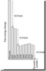
5. The free energy diagram of Glycolysis shown in Figure. What do you confer or propose from this. 6M

6. What is the difference between climacteric and non-climacteric fruit? Give three examples? 6M

7. What is "photorespiration" and why it is needed by plants? 3M

8. How is the depletion of ozone affecting the plankton levels in the oceans? 3M

9. Why virulence lost on sub-culturing? 6M

10. What are abzymes and ribozymes. Explain the differences and similarities? 6M?

Thanks for HelpBiotech

Some other enzymatic tools in rDNA technology

Polynucleotide Kinase:

  • It is a phosphorylating enzyme that transfers the gamma phosphate of ATP to a dephosphorylated end of DNA or RNA.
  • The enzyme is encoded by a gene of phage T4 and is extracted from E.coli cells infected with the phage. 
  • This enzyme used after the Alkaline Phosphatase activity and introduces 32P label (by using ATP).of DNA and RNA strands.
  • Mg+2 and dithiothreitol are used in the reaction.

Terminal Deoxynucleotidyl Transferase

  • The enzyme is a DNA polymerase that extend a strand without using a template.
  • Any nucleotide that is provided in the reaction mixture is utilized to elongate the DNA strand.
  • If only one kind of nucleotide is provided a mononucleotide polymer will be produced.

Alkaline Phosphatase

  • The enzyme alkaline phosphatase removes the phosphate moiety at the 5’-end of DNA strand, whether it is part of blunt end single extension or a recessed end of a double-stranded DNA.
  • The phosphate of RNA terminal is also removed by this enzyme.
  • The commercial source of this enzymes is two sources: Bacterial and Calf intestinal phosphatases.

RNA dependent DNA polymerase:

  • RNA dependent DNA polymerase is Reverse Transcriptase (RT).
  • This enzyme synthesizes a single strand of DNA along an RNA template.
  • It can also synthesize a second strand along the first one to make a ds complementary or cDNA.
  • RT is usually utilized to copy mRNAs into ss or ds cDNA, and to make short labeled probes.

RNase H:

  • RNase H is an endonuclease that is useful for degrading the RNA strand from a DNA:RNA hybrid molecule. It cut up the RNA into short fragments.

DNA ligases are chemical switchers

The complimentary ends of the DNAs specifically associate under annealing conditions and are covalently joined through the action of an enzyme named DNA ligase. The enzyme produced by bacteriophage T4.

Mechanism of DNA ligase activity:

The E.Coli and T4 ligases share the property of sealing nicks that have 3'’-OH and 5’- P termini. Both enzymes under take a two step reaction, involving an ‘enzyme-AMP complex’.

ligase


  • The E.Coli and T4 enzyme use different cofactors.
  • The E.Coli enzymes uses NAD as a cofactor, T4 enzyme uses ATP.

The AMP of the enzyme complex becomes attached to the 5’-Phosphate of the nick; and then a phosphodiester bond is formed with the 3’-OH terminus of the nick, releasing the enzyme and the AMP.

Special Vectors for Cloning

Some of the special vectors are using for gene cloning purpose (recombinant DNA technology). they are

a) Ti-Plasmids

b) Shuttle Vectors

c) Expression vectors

a) Ti-Plasmid:

The DNA segment, which is transfected is called “T-DNA” and is part of a large “Ti-plasmid”(Tumor inducing), found in virulent strains of Agrobacterium tumafaciens. Similarly “Ri” (Root inducing) megaplasmids are found in the virulent strains of Agrobacterium rhizogenes. The Ti- and Ri –plasmids inducing “Crown gall disease” and “Hairy root disease”.

Structure of Ti-Plasmid:

Most Ti-plasmids have FOUR regions, which are given.

image

i) Region- A:

  • It contains “T-DNA”, which is responsible for tumor induction so that mutations in this region lead to the production of tumors with altered morphology.
  • This region transferred to plant nuclear genome, so that the region is described as “T-DNA” (Transferred DNA).

ii) Region-B:

  • Responsible for Replication

iii) Region-C:

  • Responsible for Conjugation

iv) Region-D:

  • Responsible for “Virulence”, so that mutation in this region abolishes virulence. This region is therefore called “Virulence” (vir) region” and plays crucial role in this transfer of T-DNA into the plant nuclear genome.

T-DNA (Transfer DNA):

It contains two regions:

a) ‘onc’ region”:

It contains three genes, they are “tms1”, “tms2”, “tmr”.

tms1, tms2 Representing “Shooty locus”

“tmr” representing “Rooty locus”

These genes are responsible for the biosynthesis of two phytohormones, namely, “Auxins” and “Cytokinins”. These phytohormones in their turn alter the developmental program, leading to the formation of crown gall.

b) ‘nos’ region:

    • This region responsible for the synthesis of unusual amino acid (or) sugar derivatives, which are collectively given the name “Opines”.
    • Opines are derived from a variety of compounds (eg: Arginine + Pyruvate), that are found in plant cells.
    • Two most common opines are “Octopine” and “Nopaline”.
    • These two opines synthesis responsible enzymes coding are present in “T-DNA”.
    • Outside the T-DNA, Ti-plasmid carries genes that catabolize the opines, which are utilized as a source of carbon and nitrogen.
    • The T-DNA regions on all Ti and Ri-plasmids are flanked by almost perfect “25 bp direct repeat sequences”, which are essential for T-DNA transfer.

Virulence region (vir):

The vir region (~35kbp) is organized into SIX operons, namely

Vir-A, Vir-B, Vir-D, and Vir-G is  Polycistronic

Vir-C, Vir-E  is Tumor formation

c) Shuttle Vectors:

  • Shuttle vectors are plasmids that contain replication origin sequences for two different host species.
  • The host belongs to divergent groups such as Bacteria – Yeast, Monkey – E.Coli and E.Coli – Human beings.
  • The shuttle vector carries two selectable marker genes.
  • It contains the Ori of both species and a number of markers each for the tow hosts.
  • Shuttle vectors have been designed to replicate in cells of two different species; therefore, they contain two origins of replication, one specific for each host species, as well as those genes necessary for their replication and not provided by the host cells. 
  • These vectors are created by recombinant techniques
  • Some of them can be grown in two different prokaryotic species, while others can propagate in a prokaryotic species, usually E.Coli and a eukaryotic one, e.g. yeast, plants, animals.
  • Since these vectors can be grown in one host and then moved into another without any extra manipulation, they are called shuttle vectors.
  • Shuttle vectors are have been designed to specifically satisfy this need, i.e., the initial cloning of DNA inserts in E.coli and subsequent functional tests in the species to which the DNA inserts belong. Most of the eukaryotic vectors are, in fact, shuttle vectors.
  • A shuttle vector designed to replicate in E.coli and streptomyces has been constructed as follows: 
  • 1) The modules for DNA replication in streptomyces and methylenomycin A resistance are derived from a streptomyces plasmid and  2) The replication module for maintenance in E.coli and a gene for antibiotic resistance are taken from an E.coli plasmid.

d) Expression Vectors:

  • Expression vectors contain expression signals that best transcribe/translate the foreign gene in a heterogeneous system.
  • Sometimes these signals are Promoters and Terminators of E.Colior phage genes that are added at appropriate sites on either side of the gene to be expressed.

Wednesday, December 15, 2010

Colony Hybridization

This technique is used to identify those bacterial colonies in a plate which contain a specific DNA sequence.

  • The recombinant bacterial cells are plated onto a suitable agar plate; this is the “Master plate”.
  • The sterilized vector cloth containing block cork are lowered into the master plate till the velvet touches all the colonies; the block is withdrawn and gently lowered onto the nitrocellulose filter, the bacterial cells sticking on to the velvet are transferred onto the filter.
  • A reference point is marked both on the master plate and the on replica plate to facilitate later comparisons.
  • The nitrocellulose filter is removed from the agar plate and treated with “Alkali” to lyse the bacterial cells. This also denatures the DNA released from these cells.
  • The filter is treated with “Proteinase.K” to digest and remove the proteins; the denatures DNA remains bound to the filter.
  • The filter is now backed at 800C to fix the DNA; in this the DNA-print in bacterial colonies in the same relative positions in the master plate.


image

  • The filter is now hybridized with the radioactive probe (the probe represents the sequence of DNA segment used for transformation). The unhybridized probe is removed by repeated washing.
  • The colonies, whose DNA hybridizes with the probe are detected by “Auto radiography”, only these colonies show up in the autoradiography.
  • The positions of colonies showing up in the autoradiography are compared with the master plate to identify these colonies; these colonies contain the DNA segment. The colonies are then picked up for further studies.

See the Video Tutorial

Colony Hybridization

Monday, December 13, 2010

southern blotting technique

The name of this technique is derived from the following:

  • The name of its inventor, E.M.Southern
  • The DNA-DNA hybridization that forms its basis.

It is also called “Southern blotting”, since the procedure for transfer of DNA from the gel to the nitrocellulose filter resembles blotting.

DNA sample is first digested with a restriction enzyme and digested sample is gel electrophoresed. The DNA bands in the gel are denatures into single strands with the help of an alkali solution.subsequently, the gel is laid on top of a buffer saturated filter paper, placed on a solid support, with its two edges of the filter paper immersed in the buffer. A sheet of nitrocellulose membrane is placed on top of the gel and a stack of many papers on the top of this membrane. 500 grams weight placed on the top of paper towels.

The buffer solution moves, due to capillary action, from the bottom filter paper through the gel carrying with it the denatured DNA present in the gel; the DNA becomes trapped in the nitrocellulose membrane as the buffer phases through it. This process is known as “blotting” and takes several hours to complete.

While passing through the gel, the buffer carries with it single stranded DNA, which binds on to the nitrocellulose membrane, when the buffer passes through it to the paper towels. After leaving this arrangement for a few hours (or) overnight, paper towels are removed and discarded. The nitrocellulose membrane with single stranded DNA bands blotted on to it., is baked at 800C for two to three hours to fix the DNA permanently on the membrane. This membrane now has a replica of DNA bands from agarose gel, and can be used for hybridization with radioactively labelled DNA (or) RNA probe. The membrane may then be washed to remove any unbound DNA and X-ray film is exposed to the hybridized membrane to get autoradiograph.


image

For Video Tutorial Click Here

Southern Blotting Video

Monday, November 29, 2010

How to regulate UREA cycle in human body..?

The urea cycle operates only to eliminate excess nitrogen. On high-protein diets the carbon skeletons of the amino acids are oxidized for energy or stored as fat and glycogen, but the amino nitrogen must be excreted. To facilitate this process, enzymes of the urea cycle are controlled at the gene level. With long-term changes in the quantity of dietary protein, changes of 20-fold or greater in the concentration of cycle enzymes are observed. When dietary proteins increase significantly, enzyme concentrations rise. On return to a balanced diet, enzyme levels decline. Under conditions of starvation, enzyme levels rise as proteins are degraded and amino acid carbon skeletons are used to provide energy, thus increasing the quantity of nitrogen that must be excreted.
Short-term regulation of the cycle occurs principally at CPS-I, which is relatively inactive in the absence of its allosteric activator N-acetylglutamate. The steady-state concentration of N-acetylglutamate is set by the concentration of its components acetyl-CoA and glutamate and by arginine, which is a positive allosteric effector of N-acetylglutamate synthetase.

Reaction catalyzed by N-acetylglutamate synthetase

Saturday, November 20, 2010

Amino acid structure and properties online quiz

BRUSH BACK YOUR BASICS

Check out this online quiz on the structure, chemical nature, 3-letter and the 1-letter codes of the amino acids:

Amino acid Online quiz
(Just click on the above link)

Be sure that you are also able to draw the amino acids themselves and as part of a polypeptide chain.

Tuesday, November 16, 2010

Classification of Bacteriophages the viruses attacks bacterias

Classification of Bacteriophages
On the basis of presence of single or double strands of genetic material, the bacteriophages are categorized as under:

1. The ssDNA Bacteriophages
(i) Icosahedral phages = φ ´ 174, St-1, φR, BR2, 6SR
U3 and G series e.g., G4, G6, G13, G16. All are like φ ´ 174.

(ii) Helical (filamentous)
(a) The Ft group: They are F specific phages and absorb to the tip of F type sex pilus, e.g., E.coli phages (fd, fl, N13).
(b) If group: They are absorbed to I-type sex pilus specified by R factors e.g., If1, If2, etc.
(c) The third group is specific to strains carrying RF1 sex factor.


2. The dsDNA Phages
Following are the examples of dsDNA phages:
(i) T-odd phage of E.coli e.g. T1, T3, T5, T7
(ii) T-even phage of E.coli e.g.T2, T4, T6
(iii) The other E.coli phages e.g., P1, P2, Mu, φ80.
(iv) The phages of Bacillus subtilis e.g., PBSI, PBSX, PBSI, SPOI, SPO2.
(v) The phage of Shigella a e.g., P2
(vi) The phage of Salmonella e.g., PI, P22.
(vii) The phage of Haemophilus e.g., HPl.
(viii) The phage of Pseudomonas e.g., PM2.


3. The ssRNA phages
Examples of the ssRNA bacteriophages are as below:
(i) Group I : E. coli. phages such as f2, MS2, M12, R17, fr, etc.
(ii) Group II : The QP phages.

4. The dsRNA phages
Example: The φ6 bacteriophage.

Sunday, July 4, 2010

Lecture Videos - Enzymology part 1


Lecture Series on BioChemistry I by Prof.S.Dasgupta, Dept of Chemistry, IIT Kharagpur. For more details on NPTEl visit http://nptel.iitm.ac.in
Related Posts Plugin for WordPress, Blogger...