Thursday, December 23, 2010

CSIR NET Life Science December 2010 paper

CSIR NET Life Science December 2010 paper (Based on Memory


1. Pumice is the name of the most common volcanic rock that floats. It has various air bubbles and capillaries which trap air. Which statement is correct for this rock

1. Air cavities are interconnected

2. Air cavities are not connected

3. Density of rock is more than water

4. Rock is very older


2. Which of the following is responsible for ozone hole?

1. CO2                                 2. CH4

3. Chlorine                       4. NO


3. The mean salinity of sea 35 g per liter. The main cause of this observed salinity is

1. Evaporation and rainfall

2. Photosynthesis

3. Crust erosion and surface run off

4. Rivers drainage


4. Smog is due to

1. Air pollution derived from smoke and vehicles

2. More moisture in environment

3. Increase in CO2

4. Low temperature of earth surface


5. Which of the following is not a direct consequence of green house effect?

1. Increase in sea level                                 2. Rainfall

3. Tsunami                                                        4. Global warming


-----------------------------------

DOWNLOAD

-----------------------------------

December 2010 Paper

Thanks for IFAS, Jodhpur

Saturday, December 18, 2010

What is Multiple Cloning Sites?

A multiple cloning site (MCS) is a section of DNA, usually 20+ restriction sites. These restriction sites are different areas that a gene can be inserted into, and represent complimentary ends of DNA sequences that will adhere to the section of DNA to be inserted.

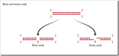
Restriction sites can have blunt, or 'sticky' ends (like an overhang), and are produced using restriction enzymes that cut the DNA in a specific way. The DNA to be inserted is then cut with the opposing sequence, so that a join can be formed.

MCS are typically found within engineered plasmids (circular double stranded DNA molecules), where they allow sections of inserted DNA be expressed.

An example of this would be the production of Insulin for sufferers of diabetes. The gene for insulin is inserted into a the MCS of a plasmid. The plasmid is then transfected into a bacteria or yeast cell, where the DNA within the plasmid is read, and expressed, leading to the production of insulin.


Few Questions about Multiple Cloning Sites:

In plasmids such as Bluescript with a multiple cloning site in a modified lacZ gene, a cloned insert can be detected by a change of colour from blue to white?

yes, the insert disrupts the lacZ gene and so if you plate the bacteria with x-gal (a substrate to turn blue), the colonies that have the insert will remain white while the colonies that don't will stay blue. lacZ when uninterrupted allows the x-gal to be metabolized and turn blue.

Why is it important that the multiple cloning site of DNA has only one restriction enzyme recognition site? I know that it makes the plasmid linear instead of circular but why does the plasmid need to be linear?

Because if it has two sites you'll end up with two pieces of DNA instead of one piece.

I have cloned my gene of interest in pUC18 at multiple cloning site. can i use this plasmid for over expression?

Yes, you can use it for over expression. Make sure your plasmid has the proper self cloning sequences, one plasmid per cell won't do you much good.
Precautions? 

Make sure your gene is pure, as in not having anything that might ruin your experiment. Test culture your target bacterium first to weed out the non functionals and then cultivate the good ones, the ones that produce your targe protein. You might need to assay for this stuff though, unless there's one of the specialized agars that will detect your target protein.

Phosphatase treated plasmid, does inhibit cloning?

Phosphatase treatment of a linearised plasmid cut in the multiple cloning site enhances vector circularization, and therefore inhibits cloning of genes into the plasmid during the ligation step.
I think that this is false, but Im not too sure why. Can you help.


it's true because phosphatase prevents vectors from closing in on themselves. This doesn't inhibit cloning but ehances it and allows a greater chance for the insert getting in the plasmid and not just the vector re-closing in on itself.

If you have any other questions about plasmids, cloning in bacteria, etc. feel free to contact me. I do this in lab almost daily

Source(s):
http://en.wikipedia.org/wiki/Multiple_cloning_site
http://www.accessexcellence.org/RC/VL/GG/transfer_and.html
http://www.genomex.com/vector_maps/pBluescript_II_KSplus_map.pdf
http://faculty.plattsburgh.edu/donald.slish/Transformation.html
http://en.wikipedia.org/wiki/Restriction_enzyme

CSIR UGC NET Paper II Model questions–part 3

1. Why proline and gylcine are exceptions in Ramachandran plot?

2. Let’s assume Hexokinase having Vmax 100 mmol/min with having Km 10 mM for Glucose.

a. With [S] = 100 mM, which will increase the velocity more; a 10 fold decrease in Km or a 10 fold increase in Vmax

b. With [S] = 10 mM, which will increase the velocity more; a 10 fold decrease in Km or a 10 fold increase in Vmax

3. A population contains 1800 individuals, 162 with yellow fruit and 1638 with green fruit. You know that fruit color segregates as a single Mendelian locus with two alleles. An experimental cross between a green-fruited and yellow-fruited individual yields only green-fruited offspring. Assuming that the population is in Hardy-Weinberg equilibrium, how many of the green-fruited individuals are homozygous at this locus?

4. Describe a strategy to sequence a 4 kb fragment of DNA.

5. Why is proline considered as a ‘compatible solute’ in plants? Mention the two molecules from which its biosynthetic pathways originate.

6. How the Pyruvate dehydrogenase (PDH), a key enzyme in citric acid cycle regulated? Name one metabolite involved in activation and one in deactivation of PDH, and mention their respective targets.

7. Show pictorially E. coli lac operon. What kind of regulation occurs in lac operon. Why?

8. a. What is popular plasma membrane model. Why is it so.

b. What are the four major phospholipids present in the plasma membrane of mammalian cells?

9. How different Cycloheximide and chloramphenicol in inhibiting protein synthesis ?

10. If you are given the genotypic frequencies of AABB, AaBB, aaBB, AABb, AaBb, aaBb, AAbb and Aabb in a mating experiment. How will you test whether the loci A and B are linked

Wish You All The Best

Thursday, December 16, 2010

CSIR UGC NET Paper II some previous - Part 2

Hope it will be beneficial.

1.  a. What is the pH of Distilled water? Can give the reasons? 3M
b. What essential is the difference between distilled and deionized water? 3M

2. Can you draw competitive and non-competitive inhibition Line-weaver-Burke Plots. Explain these different trends with respect to the mode of inhibition. 6M

3. a. Explain the condition under which two species can co-exist in a single niche? Don’t they reject competitive exclusion principle. 3M
b. Explain relationship between Biomass and productivity. 3M
c. Explain adaptive radiation with example of Darwinian finches.3M

4. a. At pH 7, polylysine, which is a polypeptide in which all the residues are lysine, adopts a random coil conformation with no regular secondary structure, However, at pH 12 it is present as an α-helix. Explain. 3M

Residue Number

Phi (deg)

Psi (deg)

1

-60

147

2

-49

-32

3

-67

-34

4

-58

-49

5

-66

-32

6

-82

-36

7

-69

-44

8

-61

-44

9

-72

-29

10

-66

-65

11

-67

-23

image

5. The free energy diagram of Glycolysis shown in Figure. What do you confer or propose from this. 6M

6. What is the difference between climacteric and non-climacteric fruit? Give three examples? 6M

7. What is "photorespiration" and why it is needed by plants? 3M

8. How is the depletion of ozone affecting the plankton levels in the oceans? 3M

9. Why virulence lost on sub-culturing? 6M

10. What are abzymes and ribozymes. Explain the differences and similarities? 6M?

Thanks for HelpBiotech

CSIR JRF/NET Life Sciences Paper II Model questions Part 1

1.  a. How many moles of CaCl2 would be used in the making of 5.00 x 102 cm3 of a 5.0 M solution?
b. Battery acid is generally 3 M H2SO4. Roughly how many grams of H2SO4 are in 400mL of this solution?
c. Why does ice float on water?

2. a. Why must cells be small?
b. The RNA is Pre-Biotic? Justify your Answer.

3. How DNA triple helix stabilized. Assume, you have isolated a E coli mutant, which contains genome as triple helix. What way it will have different fundamental processes.

4. What is Collagen and a-keratin? Compare 1°, 2°, tertiary, and quaternary structures of collagen and a-keratin.

5. How do you determine shape and size of biomolecules in solution?

6. Can you suggest three techniques used for the identification and separation of sDNA and dsDNA and give principle of each of them?


7. What factors determine variability in terrestrial ecosystem productivity?

8. How feedback loops involved in regulation stomatal opening and closing.
9. a. You have purified a RNA binding protein from E.coli cell lysate. Can you design different experiments to confirm it?
b. What is tautomeric shift in a purine of pyrimidines base? Schematically explain how tautomeric shift in a base in DNA may lead to mutation.

10. What is programmed cell death? Give important features of the PCD. Give Instances, where it occurs during Plant Development.

Note: Do not forget to say Thanks for HelpBiotech team

Thank you for HelpBiotech & its team from Bioscienceden

(source: helpbiotech)

Some other enzymatic tools in rDNA technology

Polynucleotide Kinase:

  • It is a phosphorylating enzyme that transfers the gamma phosphate of ATP to a dephosphorylated end of DNA or RNA.
  • The enzyme is encoded by a gene of phage T4 and is extracted from E.coli cells infected with the phage. 
  • This enzyme used after the Alkaline Phosphatase activity and introduces 32P label (by using ATP).of DNA and RNA strands.
  • Mg+2 and dithiothreitol are used in the reaction.

Terminal Deoxynucleotidyl Transferase

  • The enzyme is a DNA polymerase that extend a strand without using a template.
  • Any nucleotide that is provided in the reaction mixture is utilized to elongate the DNA strand.
  • If only one kind of nucleotide is provided a mononucleotide polymer will be produced.

Alkaline Phosphatase

  • The enzyme alkaline phosphatase removes the phosphate moiety at the 5’-end of DNA strand, whether it is part of blunt end single extension or a recessed end of a double-stranded DNA.
  • The phosphate of RNA terminal is also removed by this enzyme.
  • The commercial source of this enzymes is two sources: Bacterial and Calf intestinal phosphatases.

RNA dependent DNA polymerase:

  • RNA dependent DNA polymerase is Reverse Transcriptase (RT).
  • This enzyme synthesizes a single strand of DNA along an RNA template.
  • It can also synthesize a second strand along the first one to make a ds complementary or cDNA.
  • RT is usually utilized to copy mRNAs into ss or ds cDNA, and to make short labeled probes.

RNase H:

  • RNase H is an endonuclease that is useful for degrading the RNA strand from a DNA:RNA hybrid molecule. It cut up the RNA into short fragments.

DNA ligases are chemical switchers

The complimentary ends of the DNAs specifically associate under annealing conditions and are covalently joined through the action of an enzyme named DNA ligase. The enzyme produced by bacteriophage T4.

Mechanism of DNA ligase activity:

The E.Coli and T4 ligases share the property of sealing nicks that have 3'’-OH and 5’- P termini. Both enzymes under take a two step reaction, involving an ‘enzyme-AMP complex’.

ligase


  • The E.Coli and T4 enzyme use different cofactors.
  • The E.Coli enzymes uses NAD as a cofactor, T4 enzyme uses ATP.

The AMP of the enzyme complex becomes attached to the 5’-Phosphate of the nick; and then a phosphodiester bond is formed with the 3’-OH terminus of the nick, releasing the enzyme and the AMP.

Special Vectors for Cloning

Some of the special vectors are using for gene cloning purpose (recombinant DNA technology). they are

a) Ti-Plasmids

b) Shuttle Vectors

c) Expression vectors

a) Ti-Plasmid:

The DNA segment, which is transfected is called “T-DNA” and is part of a large “Ti-plasmid”(Tumor inducing), found in virulent strains of Agrobacterium tumafaciens. Similarly “Ri” (Root inducing) megaplasmids are found in the virulent strains of Agrobacterium rhizogenes. The Ti- and Ri –plasmids inducing “Crown gall disease” and “Hairy root disease”.

Structure of Ti-Plasmid:

Most Ti-plasmids have FOUR regions, which are given.

image

i) Region- A:

  • It contains “T-DNA”, which is responsible for tumor induction so that mutations in this region lead to the production of tumors with altered morphology.
  • This region transferred to plant nuclear genome, so that the region is described as “T-DNA” (Transferred DNA).

ii) Region-B:

  • Responsible for Replication

iii) Region-C:

  • Responsible for Conjugation

iv) Region-D:

  • Responsible for “Virulence”, so that mutation in this region abolishes virulence. This region is therefore called “Virulence” (vir) region” and plays crucial role in this transfer of T-DNA into the plant nuclear genome.

T-DNA (Transfer DNA):

It contains two regions:

a) ‘onc’ region”:

It contains three genes, they are “tms1”, “tms2”, “tmr”.

tms1, tms2 Representing “Shooty locus

tmr” representing “Rooty locus

These genes are responsible for the biosynthesis of two phytohormones, namely, “Auxins” and “Cytokinins”. These phytohormones in their turn alter the developmental program, leading to the formation of crown gall.

b) ‘nos’ region:

    • This region responsible for the synthesis of unusual amino acid (or) sugar derivatives, which are collectively given the name “Opines”.
    • Opines are derived from a variety of compounds (eg: Arginine + Pyruvate), that are found in plant cells.
    • Two most common opines are “Octopine” and “Nopaline”.
    • These two opines synthesis responsible enzymes coding are present in “T-DNA”.
    • Outside the T-DNA, Ti-plasmid carries genes that catabolize the opines, which are utilized as a source of carbon and nitrogen.
    • The T-DNA regions on all Ti and Ri-plasmids are flanked by almost perfect “25 bp direct repeat sequences”, which are essential for T-DNA transfer.

Virulence region (vir):

The vir region (~35kbp) is organized into SIX operons, namely

Vir-A, Vir-B, Vir-D, and Vir-G is  Polycistronic

Vir-C, Vir-E  is Tumor formation

c) Shuttle Vectors:

  • Shuttle vectors are plasmids that contain replication origin sequences for two different host species.
  • The host belongs to divergent groups such as Bacteria – Yeast, Monkey – E.Coli and E.Coli – Human beings.
  • The shuttle vector carries two selectable marker genes.
  • It contains the Ori of both species and a number of markers each for the tow hosts.
  • Shuttle vectors have been designed to replicate in cells of two different species; therefore, they contain two origins of replication, one specific for each host species, as well as those genes necessary for their replication and not provided by the host cells. 
  • These vectors are created by recombinant techniques
  • Some of them can be grown in two different prokaryotic species, while others can propagate in a prokaryotic species, usually E.Coli and a eukaryotic one, e.g. yeast, plants, animals.
  • Since these vectors can be grown in one host and then moved into another without any extra manipulation, they are called shuttle vectors.
  • Shuttle vectors are have been designed to specifically satisfy this need, i.e., the initial cloning of DNA inserts in E.coli and subsequent functional tests in the species to which the DNA inserts belong. Most of the eukaryotic vectors are, in fact, shuttle vectors.
  • A shuttle vector designed to replicate in E.coli and streptomyces has been constructed as follows: 
  • 1) The modules for DNA replication in streptomyces and methylenomycin A resistance are derived from a streptomyces plasmid and  2) The replication module for maintenance in E.coli and a gene for antibiotic resistance are taken from an E.coli plasmid.

d) Expression Vectors:

  • Expression vectors contain expression signals that best transcribe/translate the foreign gene in a heterogeneous system.
  • Sometimes these signals are Promoters and Terminators of E.Colior phage genes that are added at appropriate sites on either side of the gene to be expressed.

Wednesday, December 15, 2010

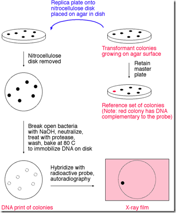
Colony Hybridization

This technique is used to identify those bacterial colonies in a plate which contain a specific DNA sequence.

  • The recombinant bacterial cells are plated onto a suitable agar plate; this is the “Master plate”.
  • The sterilized vector cloth containing block cork are lowered into the master plate till the velvet touches all the colonies; the block is withdrawn and gently lowered onto the nitrocellulose filter, the bacterial cells sticking on to the velvet are transferred onto the filter.
  • A reference point is marked both on the master plate and the on replica plate to facilitate later comparisons.
  • The nitrocellulose filter is removed from the agar plate and treated with “Alkali” to lyse the bacterial cells. This also denatures the DNA released from these cells.
  • The filter is treated with “Proteinase.K” to digest and remove the proteins; the denatures DNA remains bound to the filter.
  • The filter is now backed at 800C to fix the DNA; in this the DNA-print in bacterial colonies in the same relative positions in the master plate.


image

  • The filter is now hybridized with the radioactive probe (the probe represents the sequence of DNA segment used for transformation). The unhybridized probe is removed by repeated washing.
  • The colonies, whose DNA hybridizes with the probe are detected by “Auto radiography”, only these colonies show up in the autoradiography.
  • The positions of colonies showing up in the autoradiography are compared with the master plate to identify these colonies; these colonies contain the DNA segment. The colonies are then picked up for further studies.

See the Video Tutorial

Colony Hybridization

Tuesday, December 14, 2010

Applications of Polymerase Chain Reaction

Applications of PCR:

  • PCR can be used to amplify a specific gene in different individuals of a species. These copies can be used for cloning.
  • PCR can be used to study & identify the multiplication changes in the amplified (or) clones gene.
  • PCR can be used to study “DNA polymorphism” in the genome using known sequences as primers.
  • PCR can be used to identify the transgenic animals among the normal animals and detect the presence of a gene transferred into an organism (transgene).
  • In another hybridization methods (Southern, Northern & Western) radioactive atoms will use for identify transgene.  But by using PCR no need to radioactive atoms(P32) for transgene identification. This PCR detection takes in one day for completing this identification.
  • Use to estimate the DNA frequencies in the species.
  • PCR use to determine the physical location of genes in chromosomes.
  • By using PCR, to determine the sex of embryos.

Principle and Procedure of PCR

See the Video Tutorial of the PCR

PCR Tutorial

Monday, December 13, 2010

southern blotting technique

The name of this technique is derived from the following:

  • The name of its inventor, E.M.Southern
  • The DNA-DNA hybridization that forms its basis.

It is also called “Southern blotting”, since the procedure for transfer of DNA from the gel to the nitrocellulose filter resembles blotting.

DNA sample is first digested with a restriction enzyme and digested sample is gel electrophoresed. The DNA bands in the gel are denatures into single strands with the help of an alkali solution.subsequently, the gel is laid on top of a buffer saturated filter paper, placed on a solid support, with its two edges of the filter paper immersed in the buffer. A sheet of nitrocellulose membrane is placed on top of the gel and a stack of many papers on the top of this membrane. 500 grams weight placed on the top of paper towels.

The buffer solution moves, due to capillary action, from the bottom filter paper through the gel carrying with it the denatured DNA present in the gel; the DNA becomes trapped in the nitrocellulose membrane as the buffer phases through it. This process is known as “blotting” and takes several hours to complete.

While passing through the gel, the buffer carries with it single stranded DNA, which binds on to the nitrocellulose membrane, when the buffer passes through it to the paper towels. After leaving this arrangement for a few hours (or) overnight, paper towels are removed and discarded. The nitrocellulose membrane with single stranded DNA bands blotted on to it., is baked at 800C for two to three hours to fix the DNA permanently on the membrane. This membrane now has a replica of DNA bands from agarose gel, and can be used for hybridization with radioactively labelled DNA (or) RNA probe. The membrane may then be washed to remove any unbound DNA and X-ray film is exposed to the hybridized membrane to get autoradiograph.


image

For Video Tutorial Click Here

Southern Blotting Video

Sunday, December 12, 2010

Polymerase Chain Reaction(PCR)

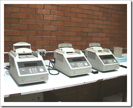
The polymerase chain reaction (PCR) technique was first developed by “Kary Mullis” in 1985,. By using the PCR technique. We can multiply (or) increases the DNA from microgram (mg) level to the higher amount level. The PCR process doing by the machines, they are “Thermocyclers”.

image

The PCR is carried out in vitro ( in test tube). It utilizes:

  • Desired DNA fragment
  • Two nucleotide primers (about 20 bases long)
  • The four deoxynucleoside triphosphates E.g.: TTP, dCTP, dATP, dGTP
  • Heat stable DNA polymerase E.g.: Taq polymerase (isolated from bacterium the “Thermus aquaticus”), Pfu polymerase (isolated from “Pyrococcus furiosus”)

Procedure:

Before starting PCR, reaction mixture will be prepare. It contains amplifable DNA, excess of the two primer molecules, four deoxyribonucleoside triphosphates and DNA polymerase.

Step 1:(Denatureation step): The reaction mixture is heated to a temperature (usually 90 to 980C) that assures DNA denaturation.

Step 2:(Annealing step): The mixture is now cooled at a temparature (40 to 600C) that permits annealing of the primer to the complementary sequences in the DNA. These sequence locate at 3’ ends of two strands of the desired segments

Step3: The temparature adjusts for the primer extending process through the 3’-hydroxyl group of primers (only annealed primer molecules). The primers are extended towards each other so that the DNA segment lying between the two primers is copied . In this polymerization “Taq polymerase” enzyme plays an important role in the polymerization process in the replication in vitro. The completion of the step3 completes the first cycle of amplification each cyle may take place one to three minutes.

Step 4: The next cycle of amplification is initiated by denaturation, which seperates the DNA strand from newly synthesized complementary DNA strand.

Step 5: Annealing allows the primers to base pairs with both the new and old strands, the totla number of strands being twice their oiginal number.

Step 6: Synthesis of new strands takes place, which doubles the number of copies of the desired DNA segment present at the end of step1. This completes the second cycle.

image

Thus at each cycle, both new and old strands anneal to the primers and serve as emplates for DNA synthesis. The end of ‘n’ cycles 2n copies of the segment are expected. The cycle may be repeated upto 60 times, but usually 20 to 30 cycles are adequate.

After PCR cycles, the amplified DNA segment is purified by gel electrophoresis and can be used for the desired purpose.

Saturday, December 11, 2010

Lodish’s Molecular Cell Biology 6e


Lodish Molecular Cell Biology, 6eimage

ISBN 10: 1429203145

ISBN 13: 9781429203142

Publisher: W.H.Freeman & Co Ltd
Publication Date: 2007
Binding: Hardcover


Synopsis:

Life Begins with Cells - Chemical Foundations - Protein Structure and Function -

Basic Molecular Genetic Mechanisms - Molecular Genetic Techniques -

Genes, Genomics, and Chromosomes -

Transcriptional Control of Gene Expression -

Post- transcriptional Gene Control -

Visualizing, Fractionating, and Culturing Cells -

Biomembrane Structure -

Cellular Energetics -

Moving Proteins into Membranes and Organelles -

Vesicular Traffic, Secretion, and Endocytosis -

Cell Signaling I: Signal Transduction and Short-term Cellular Responses -

Cell Signaling II: Signaling Pathways that Control Gene Activity -

Cell Organization and Movement I: Microfilaments -

Cell Organization and Movement II: Microtubules and Intermediate Filaments -

Integrating Cells into Tissues - Regulating the Eukaryotic Cell Cycle -

Cell Birth, Lineage, and Death -

The Molecular Cell Biology of Development -

Nerve Cells -

Immunology -

Cancer

----------------------------------------

Download the ebook

----------------------------------------

Friday, December 10, 2010

June 2010 UGC NET Life Science Previous Paper

CSIR-UGC-NET (June 2010) Life Science Previous Paper


-------------------------------
------------------------------
Do not forget to say Thanks for IAFS (Institute for Advanced Science)
Just post your comment to say thanks in this blog.

CSIR UGC NET Life Science previous papers

CSIR UGC NET life science previous papers are very useful to UGC NET aspirants
CSIR UGC NET papers for Download
CSIR UGC NET Life science Syllabus (Download)
S.No.
Year of Paper
Download link
1
2
3
4
December 2008
5
6
7
Note: Do not forget to say Thanks for IFAS (Institute for Advanced Science)

Transformation methods in animals in rDNA technology

Several approaches have been used for the introduction of DNA into animal cells/ embryos, which are listed as follows:

1) Calcium phosphate Precipitation method

2) DEAE-Dextran mediated transfection

3) Lipofection

4) Electroporation

5) Microinjection method

6) Retroviral infection method

1) Calcium Phosphate method:

clip_image002

The DNA preparation to be used for transfection is first dissolved in a phosphate buffer, calcium chloride solution is then added to the DNA solution; this leads to the formation of insoluble calcium phosphate which co-precipitates with the DNA. The calcium-phosphate DNA precipitate is added to the cells to be transfected. The cells take in the precipitate particles by “Phagocytosis”. Initially, 1-2% of the cells was transfected by this approach. In a small proportion of the transfected cells, the DNA becomes integrated into the cell genome producing stable (or) permanent transfection.

2) DEAE-Dextran mediated transfection method:

DEAE-dextran (Diethyl amino ethyl – dextran) is water soluble and polycationic i.e., has a multiple positive charge. It is added to the transfection solution containing the DNA. DEAE-dextran brings about DNA uptake by the cells through “Endocytosis”. Posing its interaction with the negatively charged DNA molecules and with the components of the cell surface plays an important role.

3) Lipofection:

clip_image004The delivery of DNA into cells using liposomes are “Lipofection”. Liposomes are small vesicles prepared from a suitable lipid. Initially, nonionic lipids were usd for preparing liposomes so that DNA had to be introduced within the vesicles following specific encapsidation procedures.

  • Usually liposomes are prepared by dispersion of a Phospholipid like Phosphotidyl choline (PC) in water by mechanical methods like Sonication, which tend to destroy DNA. DNA of up to 1 kb has been incorporated into sonicated liposomes.
  • Use of choline cationic liposomes to which DNA binds on the outside by electrostatic attraction. These liposomes cause perturbations in plasma membrane due to which they fuse and the DNA enter into the cytoplasm. Cationinc liposomes are available commercially (marketed as “LIPOFECTIN” by “Gibco-BRL”).

The positively charged liposomes not only complex with DNA, but also bind to cultured animal cells and are efficient in transforming them, by fusion with the plasma membrane. The use of liposomes as a transformation (or) transfection system is called “Lipofection”.

4) Electoporation:

Transfection mixture containing cells and DNA is exposed to very brief period (few milliseconds) to a very high voltage gradient (eg: 4000 to 8000v/cm). This includes transient pores in the cell membranes through which DNA seems to enter the cells. Linearized DNA is far more efficient in transfection than circular supercoiled DNA.

5) Microinjection:

clip_image007DNA solution is injected directly into the nucleus of a cell (or) into the male pronucleus of a fertilized one-to –two-cell ovum. The general procedure for microinjection is as follows. Donors’ females are induced to superovulated using appropriate hormone treatments. Female mice are subjected to a regime of pregnant mare serum gonadotrophin (PMSG), which stimulate growth, and development of follicles, which contain the developing oocytes. The ovulation induced by the subsequent treatment with human chronic gonadotropin (HCG). The superovulated females re then mated with fertile males, and collect fertilized eggs surgically.

The transgene construct is prepared in a buffer solution and is injected into the male pronuclei of fertilized eggs using a microinjection assembly. The linearized transgene constructs is injected into the cytoplasm of a single ovum. The microinjection embryos are cultured invitro up to the morula (or) blastocyst stage. The surviving embryos are then transferred into the uterus of surrogate mothers. These embryos develop to full term and give rise to normal mice. The microinjection method is frequently using in “transgenic biology” for producing “transgenic animals” and “Transgenic plants”.

6) Retroviral infection:

Recombinant retroviruses produce virions, which are used to infect animal cells and mice embryos. Generally, early 4 to 16 celled embryos are used for this method. The recombinant retrovirus RNA genome is copied by “Reverse transcriptase” to yield a DNA copy (reverse transcription) which becomes integrated into the cell’s genome. The reverse transcriptase is encoded by the retrovirus. And is produced immediately after infection. The recombinant retrovirus integrates into the cellular genome at random sites & usually is not accompanied with dilations (or) rearrangements.

Thursday, December 9, 2010

Ligation Methods in rDNA technology–Part 2

4) Joining with Linkers:
“Linkers are short pieces of double stranded DNA containing a restriction site. It is used to join blunt ended DNA fragments.”
In this process the linkers are attached to the blunt ends of desired DNA with a restriction enzyme, which cuts the linker to produce sticky ends. The same enzyme is used to cut the plasmid. Then the desired DNA and plasmid DNA fragments are mixed. Recombinant DNA and the linker of desired DNA.
image
5) Joining with Adapters:
Adaptor is a short DNA fragment containing one sticky end and another blunt end”
The adaptor is very similar to linkers, but it differs in having one sticky end. It is used to join blunt ended foreign DNA with plasmid DNA. The adaptors can be attached to the blunt ended foreign DNA fragment with the help of DNA ligase. As a result the blunt ended foreign DNA becomes sticky ended. It can be joined with the plasmid sticky ended DNA fragments in the normal method.

6) Alkaline phosphatase method:
By treating the liberalized plasmid vector DNA with alkaline phosphatase to remove 5’-terminal phosphate groups, both recircularization and plasmid dimmer formation are prevented. In this case, circularization of the vector can occur only by insertion of non-phosphatase treated foreign DNA which provides one 5’-terminal phosphate at each join. One nick at each join remains un-ligated but after transformation of host bacteria, cellular repair mechanism reconstitutes the intact duplex.

Ligation methods in rDNA technology–Part 1

Methods of insertion of foreign gene into the vector
There are two types of ligation of foreign gene with plasmid DNA. They are cohesive end ligation and blunt end ligation. The ligation depends upon the nature of the cutting of DNA by the restriction enzyme. The different types of insertion of foreign DNA fragment into the plasmid DNA are explained below: 

1) Cohesive end ligation:
The EcoRI cuts the DNA and produces double stranded DNA with cohessive tails. This complementary single stranded tails are often called “Sticky” mortise – and- tanon termini. This property of the enzyme was used to join both ends of a foreign gene with the ends of plasmid DNA.
The Restriction endonucleases like EcoRI, Bam HI, Sau 3A etc cut the DAN around the axis (or) at the line of symmetry of the restriction site. As a result of the cutting, linear double-stranded DNA fragments are formed; each DNA fragment has a single stranded tail at each of both strands. The single stranded ends are ready to form base pairing with each other; hence such DNA fragments are called sticky ended molecules. The same restriction enzyme is also used, to cut the foreign DNA. The complementary bases found at the single-stranded tails of foreign DNA and that those of the vector DNA undergo hydrogen bonding. But hydrogen bonding cannot seal the nick present in between the two DNA fragments. Then an enzyme DNA ligase seals the nick. As a result, the chimeric plasmid DNA is formed. The chimeric plasmid is then inserted into the bacterial cells for bacterial transformation.

2) Blunt end ligation:
Hargobind khorana (1970) first discovered the use of T4-DNA ligase in gene cloning. The blunt end ligation is practiced when Restriction endonucleases cuts the two strands of DNA along the line of symmetry of their restriction sites. Such enzymes produce blunt ended DNA fragments when the DNAs contained in the solution is treated with the restriction enzyme. The DNA fragments do not have sticky ends for ligation. In such cases, both types of DNAs are separately treated with the restriction enzyme, which produces blunt-ended DNA fragments. Then the two types of DNA fragments are mixed together to induce ligation. The ligation reaction is catalyzed by a special group of enzyme known as “T4-DNA ligase”, which joins the blunt-ended molecules. The enzyme links the ends of the foreign DNA fragments. Phosphodiester bond is established between the 3’-hydroxyl group of one fragment and the 5’-phosphate group of another DNA fragment.
image
Disadvantages:
1) A large number of plasmids are re-circularized by the action of T4-DNA ligase enzyme, the enzyme even links the two ends of the same DNA fragment.
2) The percentage of formation of recombinant plasmids containing the foreign DNA is less than that of homopolymer tailing technique of insertion of foreign gene.

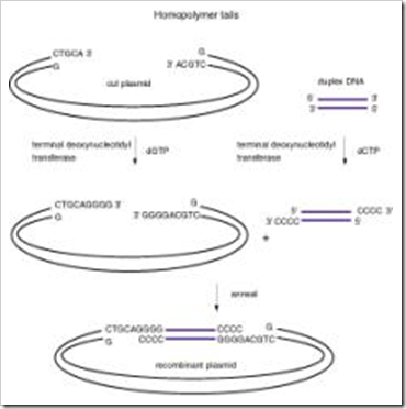
3) Homopolymer tailing:
“P.Lobban” and “Kasier” introduced this method. In this method also the foreign DNA and the plasmid are separately treated with a restriction enzyme to cut DNA into fragments. But this method need not require to produces cohesive ended (or) blunt ended molecules are used to cut the at their restriction site. The enzyme “Terminal nucleotide transferase” is used to add nucleotides to the 3’-hydroxyl group of the DNA fragments. It is a special kind of “polymerase” enzyme, which does not require template strand to add nucleotides to 3’-Hydroxyl group end of the DNA fragments. But the exact sequence of the nucleotides added to the 3,-hydroxyl group depends upon the kind of nucleotides available in the pool.
image
 
In this method, both plasmid DNA and foreign DNA are separately treated with a restriction enzyme to produce linear DNA fragments. Then the plasmid DNAs are treated with “Terminal nucleotide transferase” enzyme in presence of ATP molecules. The enzyme adds Adenine nucleotides to the DNA results in the formation of polyadenine tail [Poly (A) tail] at 3’-hydroxyl group end of plasmid DNA fragment.
On the other hand, the foreign DNA fragments are treated with terminal nucleotide transferase in the presence of TMP nucleotides; the enzyme adds thymine nucleotides to 3’-Hydroxyl group of foreign DNA fragments, forms poly thymine tail (poly T) at 3’-hydroxyl end of DNA fragment.
image
The two kinds of DNA fragments are then mixed together in a solution to establish the insertion of foreign DNA into the plasmid DNA. Complementary base pairing carries this out. The DNA ligase is used to seal the nick found in between the two fragments. 

Properties of a Good Host

Change that a normal cell undergoes as it becomes malignant; also, permanent, heritable alteration in a cell resulting from the uptake and incorporation of foreign DNA into genome.

In organisms like bacteria and other microbes, (or) even in higher plants, the uptake of genes by cells is often described by the term “Transformation”. However in animals this term has been replaced by the term “transfection”, because the term “Transformation” in animal cell culture is used to describe phenotypic alteration of cells.

Properties of a Good Host

A good host should have the following features:

(1) is easy to transform,

(2) Supports the replication of recombinant DNA,

(3) is free from elements that interfere with replication of recombinant DNA,

(4) Lacks active restriction enzymes, e.g., E. coli K12 substrain HB 101,

(5) does not have methylases since these enzymes would methylate the replicated recombinant DNA which, as a result, would become resistant to useful restriction enzymes, and

(6) is deficient in normal recombination function so that the DNA insert is not altered by recombination events.

Wednesday, December 8, 2010

Lehninger's Principles of Biochemistry, 4th edition

The Lehninger text has a long history, but given that biochemical knowledge doubles every 5 years or so, it matters what a text offers now, not in the past. The writing is simple, direct, engaging, not too easy but neither too esoteric.

The principles (as the title suggests) and the unity in diversity are emphasized, so that the student understands biochemical principles not merely facts, acronyms, pathways.

The graphics are very professional. They are comparable to any review article in hot journals such as Nature, Science, Cell, etc. The rendering of protein surfaces, and the different angles through which a structure is seen is outstanding (a good example is the section on the ribosomes).

The structures have been rendered from the PDB (protein data bank) coordinates. Most are rendered in the ribbon representation, but in many cases the surface is rendered in grey, depending on the level of detail.


The Lehninger pages on the most important protein folds, for example, are very helpful in giving the student a feel for the fold, the domain composition, the size, and names of model proteins one is expected to encounter over and again in the research literature. The text contains brief solutions to all the end-of-chapter problems


Click here to download

Download with this link
Lehninger Principles of Biochemistry (Nelson, W[1]. H., Freeman, 4th Ed, 2004).pdf

Tuesday, December 7, 2010

What are Phagemids ?

Phagemids are prepared artificially by combining features of phages with plasmids as the name suggests. One such phagemid, which is commonly used in molecular biology laboratories, is “Blue script II KS”, which is derived from PUC19, and is 2961 bp long. The KS designation indicates the “Orientation of poly linkers”, such that the transcription of lac Z gene precedes from the restriction site for Kpn I to that for Sac I.

It may be noted that it has the following features:

  • A multiple cloning site (MCS) flanked by T3 and T7 promoters to be read in opposite directions on the two strands.
  • An inducible Lac promoter (Lac I), upstream of Lac Z region, which complements with E.coli (lac Z) and provides the facilities for selection of chimeric vector DNA (recombinant DNA) using the criterion of white colonies (as against blue colonies obtained if no foreign DNA is inserted).
  • f(+) and f(-) origins of replication derived from a filamentous phage for recovery of sense (+) and anti sense (-) strands of lac Z gene, when host is coinfected with a helper phage.
  • An origin of replication (Col EI ori) derived from plasmid, and used in the absence of helper phage.
  • A gene for amphicillin resistance for antibiotic selection of chimeric vector.

What is Cosmids?

Cosmids is a hybrid DNA formed by the joining of a plasmid and lambda phage DNA carrying a “cos site” in brief cosmid is a plasmid carrying the cos site of a l phage DNA. The cosmid is not naturally found in living cells. It is a “constructed vector”.

Example: Col E1 cosmid is a typical cosmid used in genetic engineering.

Construction of Cosmids:

col E1 cosmid is constructed from col E1 plasmid and l-phage DNA. The plasmid is cut with a restriction endonuclease enzyme, which removes a portion of DNA from the plasmid. The same restriction enzyme is used to cut the l-phage DNA to get a DNA fragment containing “Cos site”.

These two DNA fragments are mixed together in the presence of the enzyme DNA ligase, which link together the two DNA fragments end to end. The resulting recombinant plasmid is called “Col E1 cosmid”.

Characteristic features:

  • The cosmid is a plasmid containing cos site.
  • It is a circular double stranded DNA.
  • H contains complementary single strand regions, the complementary single strand region is abbreviated as “Cos site”.
  • The cos site consists of two complementary single strands held  together by complementary base pairing both these two strands.
  • At the cos site, 3’-end of each of the DNA strands does not establish covalent bond with 5’-end of the same chain that is a definite nick is present in each of the two strands.
  • The nicks are restrained in the cosmid for a number of generations.
  • The cosmid DNA does not code for the synthesis of viral proteins.
  • The cosmid does not participate in the multiplication of phage particles.
  • The cosmid DNA packed with in the protein coat of bacteriophage. Thus the transformed virus particle is formed.

Advantages:

  • The cosmids transfers a somewhat larger foreign gene into the bacterial cell.
  • The cosmid pickup even long sized genes. Hence it is used in the genome of the organisms.
  • The cosmids are also used in the study of some non-sense sequences found in the genome of the organisms.

Disadvantages:

  • Each cosmid requires two cos sites for the successful packing of recombinant cosmid within the protein coat of bacteriophage. In the recombinant cosmids the packaging enzyme fails to pack the DNA into the protein coat of bacteriophage.
  • The transfer of gene from the transformed cells is difficult the cosmids need additional work for this gene transfer.
  • The package fails when the distance of separation exceeds 54,000 bps (or) when it is less than 38,000 bps.
  • Main use is gene cloning for successful result.

Vectors are carrier molecules using in rDNA technology

Introduction:

The DNA fragment (or) the gene of interest can be linked to a carrier molecule, which can transport the gene of interest into the host cell. This carrier molecule is referred to as a “Cloning vector” (or) “Cloning vehicle”; the cloning vehicle is the central component of a gene cloning experiment and it constitutes the gene transfer system.

Important features of vectors:

  • It must be able to replicate.
  • There must be some way to introduce vector DNA into a cell.
  • There must be some means of detecting its presence, preferably by a plating test.
  • It should contain an assortment of unique Restriction endonucleases cleavage sites.
  • It should occur in large number of copies.

Type of vector:

There are three main types of vectors in use. They are

1) Plasmids

2) Cosmids

3) Phagemids

1) Plasmids:

Plasmids are extra chromosomal, autonomously replicating , small circular molecules of DNA found in many prokaryotes and in a few eukaryotes such as the yeast “Saccharomyces cerevisiae”.

Properties of Plasmids:

  • They replicate independently (or) autonomously
  • Most of them are circular duplex of DNA molecules.
  • They have an origin of replication naturally in them
  • They are passed on to the daughter cells during cell division.
  • They may carry very important genes for antibiotic resistance, toxin production, for antibody production, for degradation of a large number of unusual substrates such as herbicides (or) industrial effluents and genes for nitrogen fixation. These confer the phenotypic traits of plasmids. 
  • They rely on the DNA replication enzymes of the host cells for their replication; however, the initiation of replication is controlled by plasmid genes.
  • Certain plasmids do not show any phenotypic traits such as plasmids are called “Cryptic plasmids”.
  • They have high transformation efficiency.
  • They have convenient selectable markers such as antibiotic resistance, toxin production etc, for transformants and recombinants.
  • They have the ability to clone reasonably large pieces of DNA say about 5 kilo base pairs.
  • They are of low molecular weight.
  • They are easily isolated and purified.

Size of plasmids:

Plasmids are duplex, supercoiled DNA molecules and they range in size from 1X10⁶ Daltons to greater than 200X106.

Number of plasmids:

The number of copies of plasmid in a cell is referred to as “Copy number”. When there are one (or) two copies, the copy number is called “low copy number”. When there are twenty (or) more copies per cell, the copy number is called “high copy number”

Plasmid classification:

The naturally occurring plasmids are classified based on the main characteristics coded by the plasmid genes. It is grouped into FIVE main types:

a) F-plasmids

b) R-Plasmids

c) Col – plsamids

d) Degradative plasmids

e) Virulence plasmids

a) F-Plasmids (Fertility plasmids):

These plasmids carry only “tra” genes (transfer gene) and no characteristic beyond the ability to promote conjugate transfer of plasmids. The presence of “tra genes” promotes bacterial conjugation. These plasmids may be denoted as F+ and F-, which means those having the fertility (F) factor and those without it. These plasmids are not used in gene cloning. Most of the “tra genes” are involved in “pili synthesis” (sex pili) on donor.

b) R-Plasmids (Drug resistance):

These carry genes conferring on the possessor resistance to one (or) more antibacterial agents such as “Chloramphenicals”, “Amphicillin”, “Tetracycline” and any metal. The “R” strands for “Resistance”. “Plasmid RP4” found in pseudomonas is an example of R-plasmid. This R-factor was discovered in Japan in 1955.

The R-factor is wide spread in contain strain of almost all pathogenic bacteria. The plasmid genes after encode for enzyme that chemically inactivate the drug (or) by active export eliminate it from the cell.

c) Colicinogenic (or) Col plasmid:

Col plasmids are E.coli plasmid able to produce colicins, proteins that prevent growth of susceptible bacterial strains that do not contain a col plasmid. The bacterial toxins are generally called “Bacteriocin” these bacteriocin are active only against closely related strains of bacteria toxins of this. Types that are liberated by strains of E.coli are called “Colicins”. The colicins are simple proteins. Several different types of colicins have been isolated which kill sensitive cells by different mechanisms. The plasmids containing genes for such toxic substance, “colicin” is called “Col plasmid”. Depending upon the nature of colicins, there are different types of col plasmids. They are col B, colE1, col E2, col I and col V. the toxin from “Pseudomonos” is called “Pyrocins”.

d) Degradative plasmids:

These are plasmids, which have genes for enzymes that enable the bacterium to metabolize unusual substrates such as Toluene, xylene and salicylic acid. These plasmids are also called “Dissimilation plasmids”. This plasmid type (Ptol) is responsible for the ability of certain Pseudomonas species to break down different to degrade industrial solvents such as toluene and xylene. A combination of several plasmids, when transferred to pseudomonas bacteria, allows the bacteria to break down complex hydrocarbons and other compounds present in crude oil. The bacteria, containing these plasmids have a potential use for treatment of environments contaminated with oil spills.

e) Virulence plasmids:

The plasmids have genes that confer pathogenicity on the host bacterium. For example, Ti-plasmids found in “Agro bacterium tumefacience”. They include crown gall disease on dicotyledonous plant.

2) Cosmids

3) Phagemids

Friday, December 3, 2010

Restriction endonucleases are chemical knives

The Restriction endonucleases recognizes a specific base sequence of four to eight bases in double-stranded DNA and cleaves both strands of the duplex.
There are known type of restriction endonucleases : type I,II and III. Type II enzymes are frequently used in the rDNA technology. Type I and type III are not use because these type enzymes cleaves the DNA far from the recognition sites. The Restriction endonuclease recognizes particular specific sequences are 4 to 6 nucleotides recognize by the type II enzymes because these enzymes only cleave at this site. The palindrome sequence is “Two-fold symmetry” as shown below.
 
image
The type II restriction rnzymes are discovered and characterized by “Hamilton Smith” and “Daniel Nathans” in 1960. Approximatly 200 Restriction endonucleases are isolated from bacterial species like- E.coli, Bacillus, Haemophillus, Streptococcus and thermus aquaticus.
Restriction endonucleases is named by the first letter of the “genus” of the bacterium that produced it and the first two letters of its “species”, followed by its serotype (or) strain designation, if any, and a roman numerical if the bacterium contains more than one type of restriction enzymes.
image
E.g.: EcoRI is produced by “E.coli” strain RY13
Bam HI isolated from “Bacillus amyloliquefaciens
Bgl II isolated from “Bacillus globigis
Hae III isolated from “Haemophilus aegyptius
Pst I isolated from “Providencia stuartil

Tool in Recombinant DNA Technology

Enzymes involved in rDNA technology:

Genetic engineering means creation of new DNA, for the cloning purpose. In these rDNA technology two types of enzymes are using:

1) Restriction endonucleases

2) DNA ligases

3) phosphatases

4) Reverse transcriptase

5) Polynucleotide kinases

6) Terminal transferase

7) Nucleases-S1 and

8) RNAase H

Thursday, December 2, 2010

What is Biotechnology..?

Some of the available definitions of Biotechnology,

 “It is the application of biological organisms, system (or) process to manufacturing and service industries”

“ The integrated use of Biochemistry, Microbiology and engineering sciences of the capabilities of microorganisms, cultured tissue cells and part of them.”
“The controlled use of biological agents, such as microorganisms (or) cellular components, for beneficial use”
The last definition is brief and comprehensive and may be used by students if they have to learn only one definition.

Historical Resume:
The term Biotechnology was coined by the scientists “Carl Neuberg” in 1919.
The origin of biotechnology can be traced back to prehistoric times when microorganisms were already used for processes like “Fermentation”.

First Discovery: In 1920, “Clostridium acetobutylicum” was used by “Chaim Weizmann” for converting starch into butanol and Acetone. The latter was an essential component of explosives during World War-I.

Second Discovery: During Second World War (the 1940s), the production of Penicillin (as an antibiotic discovered by “Alexander Flemming” in 1929) on a large scale from cultures of “Penicillium notatum

Third Discovery: The third discovery of Biotechnology is its recent reincarnation in the form of “Recombinant DNA technology”, which led to the development of a variety of gene technologies and is thus considered to be the greatest scientific revolution of this century.

Old Vs New Biotechnology:
Fermentation by some microorganisms, formation of yogurt (curd), cheese from milk, Veniger from molasses, production of antibiotics like “Penicillin” from certain fungi, Process of baking and brewing are often included in describing what is called “Old Biotechnology

PCR (Polymerase Chain Reaction), rDNA technology, Cell culture and fusion, and bioprocessing, which became possible only through the researches in molecular biology have been described as “New Biotechnology


Bioscience Den ebooks collections

cell

The CELL
Bruce Albert

Guyton's Textbook Of Medical Physiology 11th  2005
Textbook Of Medical Physiology by Guyton & Hall, 11th Edition
harper biochem

Harper’s Illustrated Biochemistry

image

Practical Biochemistry & Molecular Biology
Wilson & Walker

image

The Essential Molecules of Life
CARBOHYDRATES
Robert V Sock

image

Introduction to Protein Structure by Branden and Tooze ebook
image

Biology ebook Complete collection

image
Molecular Biology of the Cell, 5th Edition
image
How to Study for Success
image
Schaum’s Outlines of Immunology
image
Schaum's Outlines of Biochemistry (Ebook) second edition
image
Fundamentals of Biochemistry J.L.Jain
image
Ecology by Odum
   

Note: These are all Collection of referral links only

Wednesday, December 1, 2010

Global warming hastened arrival of dinosaurs

London: Global warming 300 million years ago triggered the evolutionary burst which caused tiny lizards to grow into gigantic dinosaurs, say scientists. 

Scientists now believe that the Earth became significantly hotter during this (Carboniferous) period, turning rainforests into 'islands' surrounded by arid deserts. This climatic change caused lizards to adapt to live in drier conditions and led to the evolution of different varieties of dinosaurs, according to the Daily Mail, citing the journal Geology.

Howard Falcon-Lang of Royal Holloway, University of London, who carried out the research, revealed this global warming indirectly influenced mammalian evolution. "The global warming set in motion the process which led to the evolution of dinosaurs and produced different species which adapted to the climate in different ways," he said.

According to the research, around 300 million years ago the supercontinent of Pangea was covered by a huge rainforest and the reptiles which inhabited it were very similar. However, a period of global warming caused the forest to fragment into 'islands' of trees, allowing each population of trapped reptiles to evolve in a different way.

The world became so hot that the polar ice caps melted and vast forests grew at the North and South poles. The reptiles eventually moved out of the dwindling forests and into the arid desert lands before the dinosaurs arrived around 220 million years ago.

The research team made their findings by analyzing and carbon dating hundreds of reptile fossils from around the world and analyzing how they evolved over time.

Source: IANS

Related Posts Plugin for WordPress, Blogger...